
Can a dissolving skin patch make melanoma cells easier for chemotherapy to kill?
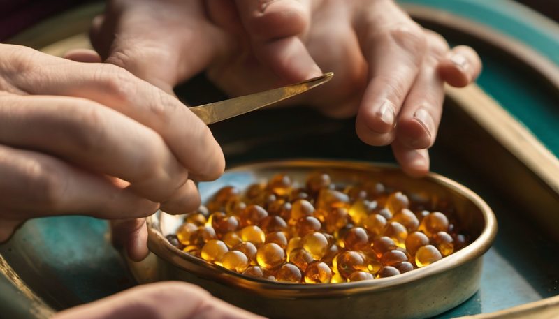
Melanoma is hard to treat because it grows, spreads and learns to ignore chemotherapy. A team in China built a dissolving microneedle patch for mice: tiny needles carrying doxorubicin and a CRISPR/Cas9 complex aimed at the YB-1 gene. The question was whether pressing such a patch onto skin could deliver both agents close enough to the tumor to change its behavior.
Quick takeaways
A needle array that dissolves where it lands
A dissolving microneedle patch is built from a grid of tiny pyramidal tips, each about 800 micrometers tall. Those tips are made from chondroitin sulfate, a sugar-like material that is biocompatible and breaks down in the body. That same material also coats the drug-carrying nanoparticles and is meant to improve uptake by melanoma cells that carry CD44. Because the tips are tall enough to pass the outer skin layer, pressing the patch can place medicine near a lesion.
Those needles dissolve at body temperature, so the medicine is released where they land. That local release showed up in live-mouse imaging, where a fluorescent stand-in for chemotherapy stayed brightest at the application site. It faded faster when simpler patches carried the same dye, while the signal did not spread to untreated skin. After pressing, puncture marks appeared in mouse skin, faded within hours, and looked largely recovered by 24 hours.
The patch therefore works as a small dissolving stamp: it presses into skin, leaves its cargo behind, and then disappears.
The survival switch that helps melanoma endure
Melanoma cells can build up a survival protein that helps them grow, spread, and resist treatment. That protein also moves into the nucleus in many cancers, where high levels have been linked to worse outcomes. The buildup gives those cells a practical edge. The edge comes from keeping one survival program running while under attack. A fast-growing cell needs to divide, cope with damage, and avoid dying when treatment arrives, so the gene behind that protein becomes a useful target.
The patch turns that target into a treatment by carrying two agents in one dissolving needle array. One agent is doxorubicin, a chemotherapy drug that inserts into DNA and triggers cell death. The other is a gene-cutting complex aimed at the survival gene. The same patch uses a sugar coating to help those carriers find melanoma cells that display a matching receptor. Those coated carriers then entered most melanoma cells in dish tests and cut many of the measured target sites. There, they left nearly all treated cells unable to survive in the assay, while simpler delivery groups allowed far more growth. Their colony counts pointed the same way, leaving treated cells with far fewer surviving clusters than other carriers.
Two drugs in one bead, and what the mice showed
A single sugar-coated bead held two medicines together. One was doxorubicin, a chemotherapy drug that damages DNA inside fast-dividing cells. The other was a ready-made gene-editing complex, made from a cutting protein and a short guide RNA.
In dish tests, the coated beads put both payloads into about three quarters of melanoma cells. They also lowered the target gene in many treated cells. The sugar coat helped the beads reach cells carrying a matching surface receptor, which can improve uptake in the tested tumor cells. Compared with uncoated particles, this targeting reduced harm to normal kidney cells.
The mouse result was larger than any dish test could show. Tumors from mice treated with the dual patch weighed 92.56 percent less than untreated tumors, about one-thirteenth of their weight. Tissue slices showed fewer dividing cells and fewer blood-vessel markers. They also had lower levels of a protein that helps tumors hide from immune attack. Those changes tracked with tumor shrinkage, but they do not prove each one caused the weight loss.
A small patch, a wider test
In C57BL/6 mice with subcutaneous B16F10 melanoma tumors, the full dissolving patch left final tumor weight at 0.073 grams instead of about 0.98 grams in untreated controls, a 92.56 percent reduction. Tumor slices from those mice also showed far fewer Ki67-positive dividing cells and less CD31 blood-vessel staining, so the smaller tumors came with quieter proliferation and vessel building rather than size alone.
Local release is where the design could matter beyond one mouse model. In live-mouse imaging, the strongest fluorescent chemotherapy signal stayed at the application site for 4 to 72 hours, while hyaluronic acid and chondroitin-sulfate-only patches faded faster. That pattern fits a patch that presses two medicines into one skin site, raises the dose near the lesion, and leaves fewer organs exposed than an injection sent through the whole body.
The test still ends on day 23, after patches were pressed onto tumors for three minutes and taped in place so the needles could dissolve. The formulation was described as lowering off-target toxicity compared with less targeted carriers, but no figure gives the size of that drop. If local genetic repair keeps its precision in human skin, the next question is how many stubborn lesions it can reach.
You may also like
Health and medicine · Genetics and biotechnology
Can an ordinary phone camera read the shape of your pulse from a color-changing skin patch?

Space and astronomy
What do calcium and tin isotopes reveal about the invisible matter inside neutron stars?

Technology and computing
Cars can be tracked through surveillance cameras nobody gave access to

Health and medicine · Brain and mind